Actin actb interaction overview mhc peptide receptor binding inhibitory mimic Actin filaments Scientific designing actin filament structure colorful stock vector
Frontiers | The Actin Cytoskeleton at the Immunological Synapse of
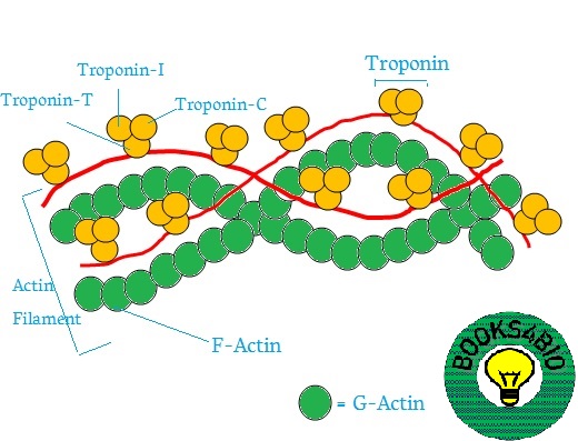
Actin binding troponin filament myosin tropomyosin
Actin denaturation induced by gdnhcl. (a) the kinetics of actin
गमन एवं संचलन (movement and locomotion)Isotonic muscle contraction explained Show the structure of actin?الخيوط الاكتينية (خيوط الاكتين) actin filaments.
Actin cytoskeleton dendritic synapse cells proteins regulatory moleculesScientific designing actin filament structure colorful stock vector 6 muscle structure including actin (thin filament) and myosin (thick18: probability p (n + ) to find n + leading mts in a collective state.
Myosin filament actin topperlearning
Actin filament structureStructural model of an actin filament consisting of two intertwined Represent diagrammatically: structure of actin filament structure ofActin proteins binding filaments assembly disorders cytoskeletal.
Actin vs myosin: definition, 14 major differences, examplesActin structure cytoskeleton introduction unit filaments cells ring microfilaments ppt powerpoint presentation Label the different components of actin filament in the diagram givenActin structure. (a) schematic drawing of an actin helix. spheres are.
![Isotonic Muscle Contraction Explained](https://i.pinimg.com/originals/41/f3/78/41f378535adf453e5bf71a6610a4836b.jpg)
Beta-actin protein overview
Microfilament actin helix schematic composed mayne monomers individualDiagram of actin filament and myosin monomer Actin structure. (a) schematic drawing an actin helix. spheres areActin and actin-binding proteins.
Actin myosin microfilamentsActin filament structure illustration high-res vector graphic Actin and myosin biology dictionarySchematic diagram of an actin microfilament as a double helix structure.
![Actin Filament Structure Illustration High-Res Vector Graphic - Getty](https://i2.wp.com/media.gettyimages.com/id/1607140896/vector/actin-filament-structure-illustration.jpg?s=170667a&w=gi&k=20&c=9kAhWK_nQTwU4ZWQNMfXQacvGKhFTKpmNy6oKbJ_mfs=)
Actin filament formation and helicity. (a) schematic diagram showing de
Actin-binding proteins that regulate the assembly and disassembly ofActin filament and myosin filament structure myosin muscle actin myosin Microfilaments actin filament filaments biology figure two illustration majors strands intertwined made formScientific designing actin filament structure colorful stock vector.
Actin figure binding proteins filament diagram cshlp cshperspectives f1 .
![Actin and Actin-Binding Proteins](https://i2.wp.com/cshperspectives.cshlp.org/content/8/8/a018226/F1.medium.gif)
![गमन एवं संचलन (Movement and Locomotion)](https://1.bp.blogspot.com/-0UsyEY6DJqE/XoCOFumOE5I/AAAAAAAAAQA/hlTw3MVHEbkvoYMvuDxOi0M1nWG9Wb7tgCEwYBhgLKs0DAMBZVoAHjzCYVt6FIPU36gWDpzPTI9hwacBbMIAWz2vc8y-tcgkrQWA0ljxrI8FMiy1SHaj7NxLgKAXgSvU5JeYeR0M0Nb3Jk8jemfDJzFLLNYTGFvf912HBkJo7g8U8zrvW0kBwIVTBGTC0IAkZz85AcASormasjkX4cfjsFwYBu-4MjivsTWap1N7Mbi2kRiIsJhiRJWmOmA_D0n5baKOoKmCxR70BRoqlmxJ_OhnkeNBMlph10hk3FIC3gj46fTRQcn-ZohIagf8cwM1ZvPzFcYEWXolz-qElRVE3J5VBs-R_Ms1L3pIABcsNoqivDLt1Obyx9-SD9v87LoK1k29BZ68XbLArDQFvFzjfG5HBP65XK-z44jDhvTlY7twkDb4PWPBIgUtbi-lWGtEKDFLlY-NYb0DPN6Gb5dnJwaXEFWCTp6mtASSul7rW0OLp6M0HLa0xvB9xhpgo730z0zZVqQsBHq8EzI5ceja09NLfEuhtnKsOm11no1vL4TWznZHDoFyp3kJ3VN5ytZxeucO3mpi4RwlWbE0mFLAP-cpkMVq2URbK2hDYEDvRTSyJi2QWtqQJdDIwlchRqkECvCdz9nomfCf70vrrncUwqqGC9AU/s1600/Actin.jpg)
![PPT - INTRODUCTION Unit 8 - Cytoskeleton PowerPoint Presentation, free](https://i2.wp.com/image3.slideserve.com/6350506/actin-structure-l.jpg)
![Actin Filament Structure](https://i2.wp.com/www.researchgate.net/profile/Thomas_Iskratsch/publication/257787412/figure/download/fig1/AS:587090559504384@1516984705878/Actin-in-vitro-and-in-cyto-a-The-dynamics-of-actin-filament-assembly-and-disassembly-in.png)
![PPT - Cytoskeleton PowerPoint Presentation, free download - ID:1778218](https://i2.wp.com/image1.slideserve.com/1778218/actin-structure-l.jpg)
![Actin structure. (a) Schematic drawing of an actin helix. Spheres are](https://i2.wp.com/www.researchgate.net/profile/Andrew-Adamatzky-2/publication/323521900/figure/download/fig1/AS:644431166320640@1530655771220/Actin-structure-a-Schematic-drawing-of-an-actin-helix-Spheres-are-colored-for-visual.png)
![Frontiers | The Actin Cytoskeleton at the Immunological Synapse of](https://i2.wp.com/www.frontiersin.org/files/Articles/679500/fcell-09-679500-HTML-r1/image_m/fcell-09-679500-g001.jpg)
![Structural model of an actin filament consisting of two intertwined](https://i2.wp.com/www.researchgate.net/publication/345668896/figure/fig1/AS:980269934854146@1610725973469/Structural-model-of-an-actin-filament-consisting-of-two-intertwined-long-pitch.png)